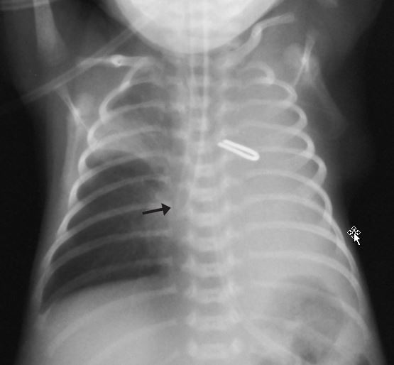
24-week premature newborn, weighing 930 grams and measuring 35cm in length, evolved with respiratory distress syndrome and patent ductus arteriosus. The infant was intubated due to low O2 saturation on the first day of life, progressing to pulmonary atelectasis. Image description: complete left hemi thorax and right upper lobe opacities. Area of increased density in the anterior segment, increased horizontal fissure and hyperinflation of the middle and lower right lobes. The tip of the endotracheal tube was positioned in the right intermediate bronchus (arrow).A metallic clip was also observed and represented clipping of ductus arteriosus.
Image description: complete left hemi thorax and right upper lobe opacities. Area of increased density in the anterior segment, increased horizontal fissure and hyperinflation of the middle and lower right lobes. The tip of the endotracheal tube was positioned in the right intermediate bronchus (arrow).A metallic clip was also observed and represented clipping of ductus arteriosus. Diagnosis
Pulmonary atelectasis of the entire left lung and right upper lobe, caused by an improperly positioned endotracheal tube. Note: In hyaline membrane disease, the radiographic picture is characterized by pulmonary opacities with reticulogranular pattern and diffuse, bilateral, peripheral air bronchograms. On x-ray, these findings were not clearly identified, as a result of atelectasis.
DiscussionHyaline membrane disease results from insufficient surfactant production, generating widespread alveolar atelectasis and respiratory distress symptoms in the first hours of life after birth. This disease is quite common in premature infants, which need to undergo assisted mechanical ventilation. Other causes of atelectasis in premature and low birth weight newborns include lung immaturity itself and malpositioned endotracheal tube. Chest radiograph should be performed to confirm clinical diagnosis of the disease and the exact location of the endotracheal tube tip. On chest x-ray, the endotracheal tube tip should be visualized at the level of thoracic vertebrae T1. If positioned below this location, primarily selective intubation of the right main bronchus may occur, obliterating the left main bronchus and causing pulmonary atelectasis. When the cannula tip is located at an even lower level in the right intermediate bronchus, the right upper lobe bronchus is also obliterated, causing complete atelectasis of the left lung and right upper lobe, as occurred in this case. In this situation, the endotracheal tube should be repositioned to resolve the obstructive process and clinical picture of pulmonary atelectasis.
Author: Mariana Chiaradia Dominguez: http://lattes.cnpq.br/2294611196717897. Fifth-year Medical Graduate Student at the Unicamp School of Medicine. She develops research projects in Neonatal Radiology in CAISM (Women’s Hospital), supervised by Prof. Dr. Beatriz Regina Alvares http://lattes.cnpq.br/3415407272085114