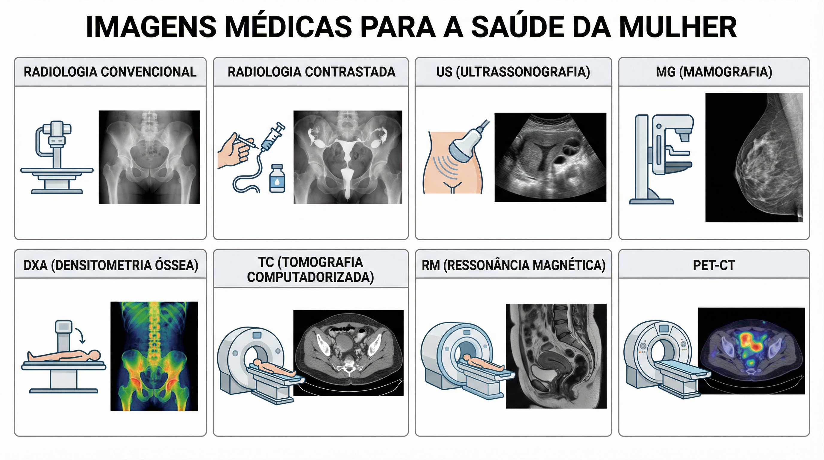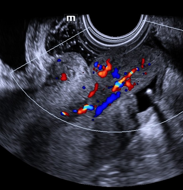
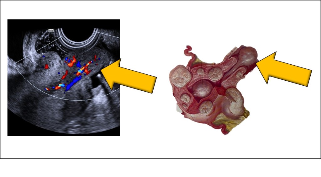
 It is observed an isoechoic to the myometrium mass, protruding from the cervical canal, toward the vagina (yellow arrows). Doppler imaging demonstrates that it is a well-vascularized mass, containing a vascular pedicle, originating from the uterine cavity. The ultrasonographic documentation of this expelled myoma was facilitated by the injection of ultrasound gel into the vaginal cavity. The anechoic texture of the gel enhances the visualization of the mass's echotexture, shape, and margins.
Conteúdo
It is observed an isoechoic to the myometrium mass, protruding from the cervical canal, toward the vagina (yellow arrows). Doppler imaging demonstrates that it is a well-vascularized mass, containing a vascular pedicle, originating from the uterine cavity. The ultrasonographic documentation of this expelled myoma was facilitated by the injection of ultrasound gel into the vaginal cavity. The anechoic texture of the gel enhances the visualization of the mass's echotexture, shape, and margins.