


Uterine spindle cell neoplasm. Magnetic resonance imaging findings.
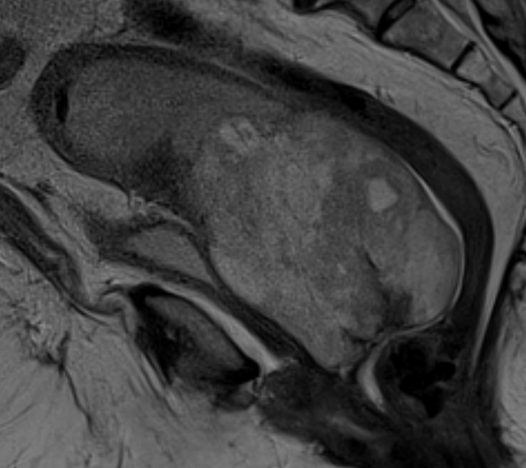
Sagital T2 mostra a mesma lesão sólida expansiva centrada no colo, com sinal alto em T2, que determina moderado hidrométrio obstrutivo, com bolhas gasosas no fundo da cavidade endometrial.
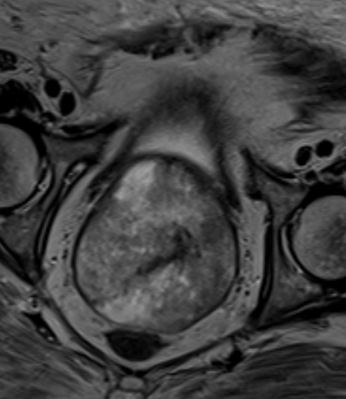
Axial T2 reveals an expansive solid lesion distending the endocervical canal, with preserved parametrium.
˗ Uterus of increased dimensions, presents a solid tumor lesion with necrotic areas in between, hypervascularized, occupying the proximal portion of the endometrial cavity and endocervical canal, measuring 10.4 x 7.5 x 8.0 cm, volume of 336.43 cm³
- The lesion determines distension of the endometrial cavity, with moderate obstructive hydrometrium (35 mm thick) and gas bubbles at the bottom of the endometrial cavity. There is loss of the endometrium-myometrium interface, inferring myometrial invasion. Note extension of the lesion into the vaginal canal. There are no signs of peritoneal extension or parametrial invasion.
- Note that this high signal on T2 is generally associated with non-epithelial neoplasms, especially sarcomatous lesions. In this case, it was a spindle cell neoplasm, a very rare diagnosis.
Graduação médica pela Universidade Federal do Rio de Janeiro (UFRJ). Residência médica em Radiologia pela UNICAMP. Título de especialista pelo Colégio Brasileiro de Radiologia. Pós-graduação em Radiologia da Mama e Pelve Feminina pelo Instituto de Radiologia da Universidade de São Paulo (InRad-USP). Médico radiologista assistente do Hospital da Mulher Prof. Dr. José Aristodemo Pinotti - CAISM/UNICAM, com atuação em Tomografia computadorizada e Ressonância Magnética.